ILLUSTRATIONS
from
MEDICINE.
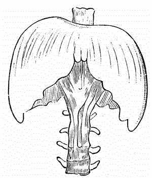
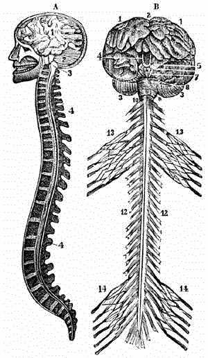 BRAIN.
A. - A section of the brain and spinal column.
B. - Anterior view of the brain and spinal cord. |
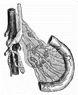
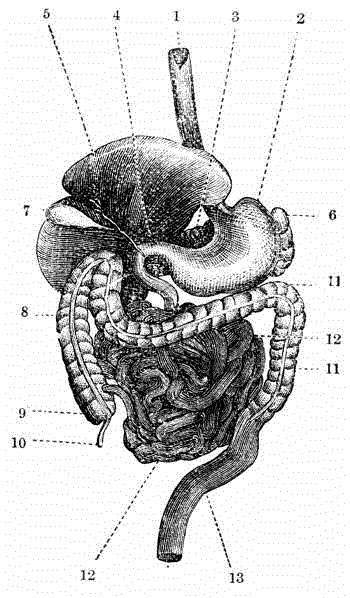 DIGESTIVE APPARATUS IN MAN. |
|
|
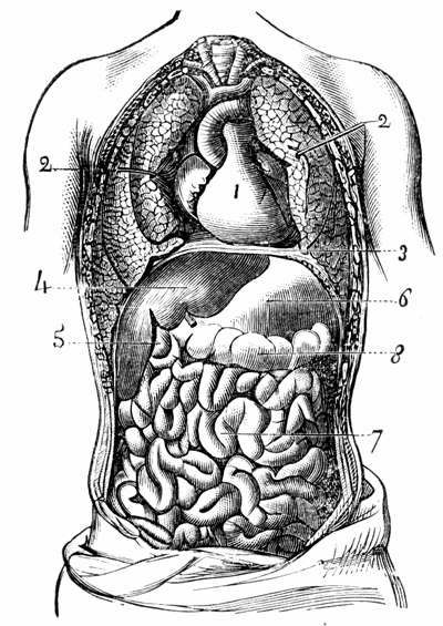
1, Gullet. 2, Stomach. 3, Pancreas. 4. Pyloris. 5, Liver. 6, Spleen. 7, Gall-bladder. |
8, Large intestine. 9, Caecum. 10, Appendix of the caecum. 11, Colon. 12, Small intestine. 13, Rectum. |
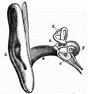 THE EAR.
|
 EXTERNAL, MIDDLE, AND INTERNAL EAR.
|
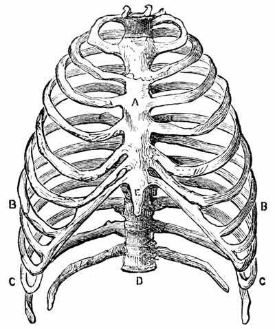 THE RIBS.
|
 ANTERIOR VIEW OF THE DIAPHRAGM IN A STATE OF REPOSE. |
 VIEW OF LARYNX FROM ABOVE.
|
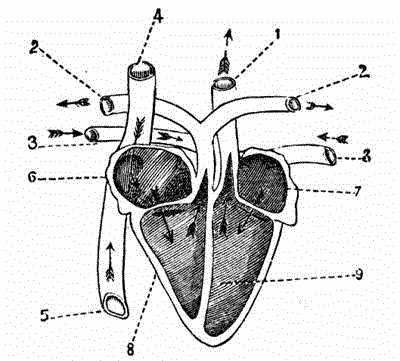 SECTION OF THE HEART.
|
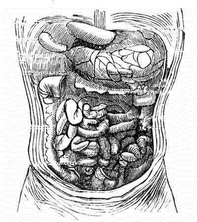 CAVITY OF THE ABDOMEN. |
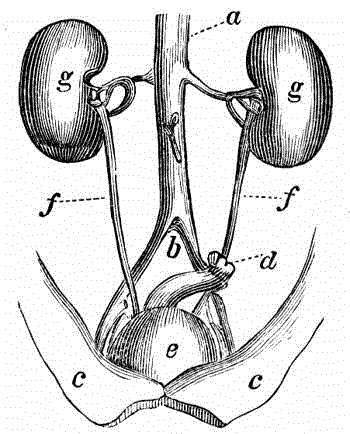 THE URETERS RUNNING FROM THE KIDNEY TO THE BLADDER.
|
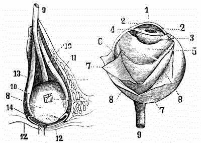
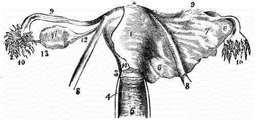 THE UTERUS AND ITS APPENDAGES VIEWED ON THEIR ANTERIOR ASPECT.
|
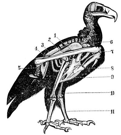 SKELETON OF THE VULTURE.
|
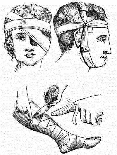 MODE OF APPLYING BANDAGES. |
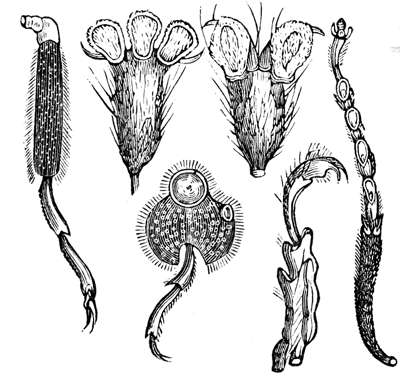 VARIOUS FORMS OF INSECTS' FEET, SHOWING THE ADHESIVE DISKS OR SUCKERS. |
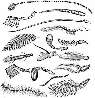 VARIOUS FORMS OF ANTENNAE. |
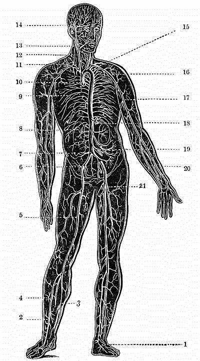 ARTERIES OF THE HUMAN BODY.
|
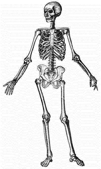 SKELETON OF MAN. |
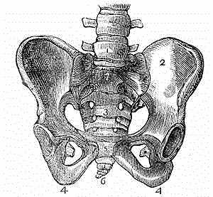 THE PELVIS.
 |
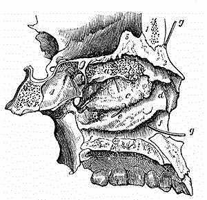 A LONGITUDINAL SECTION OF THE NASAL FOSSAE OF THE LEFT SIDE, THE CENTRAL SEPTUM BEING REMOVED. | ||
|
|
 THE SALIVARY GLANDS.
|
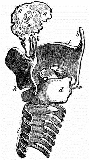
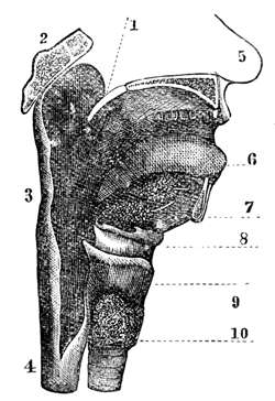 VERTICAL SECTION OF THE MOUTH AND THROAT.
|
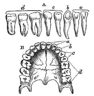 THE TEETH.
|
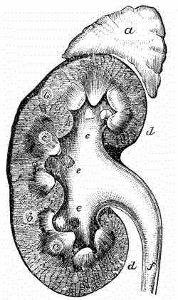 VERTICAL SECTION OF THE KIDNEY.
|
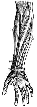
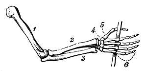 GENERAL ARRANGEMENT OF THE BONES OF THE ARM.
|
 a, b, THE CLAVICAL, OR COLLAR-BONE. |
 TRANSVERSAL VIEW OF THE THORACIC AND ABDOMINAL CAVITIES.
|
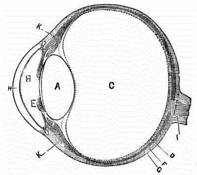
 ANATOMY OF THE EYE.
|
|
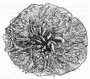 BREAST. Lactiferous ducts, dissected out and injected. |
 CAVITY OF THE EAR. |
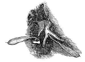
 CHYLE VESSELS OF THE MESENTERY. |
 HAND.
|
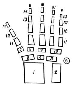 DIAGRAM OF THE BONES OF THE HAND. With the ends of the radius and ulna.
|
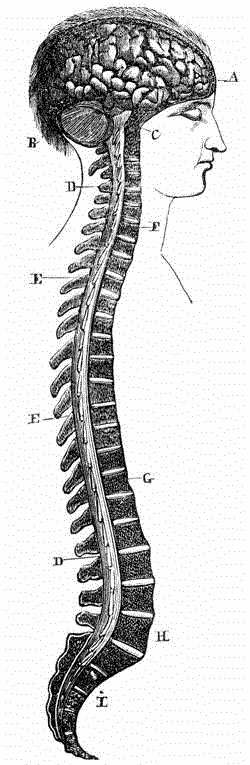 NERVOUS - CEPHALOSPINAL CENTRES.
|
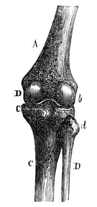 KNEE-JOINT.
|
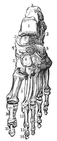 DORSAL SURFACE OF THE LEFT FOOT,
|
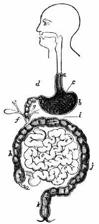 ALIMENTARY CANAL.
|
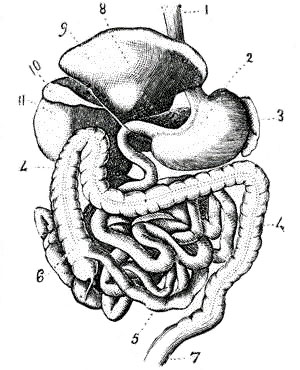 ALIMENTARY APPARATUS.
|
 CARTILAGES OF LARYNX AND EPIGLOTTIS AND UPPER RINGS OF TRACHEA, SEEN FROM BEHIND. (TAKEN FROM TODD AND BOWMAN.)
|
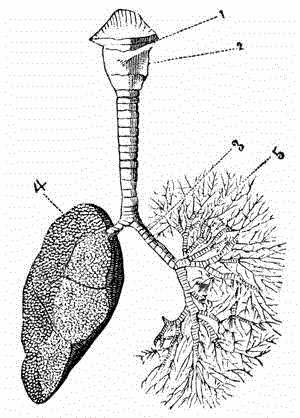 BRONCHI.
|
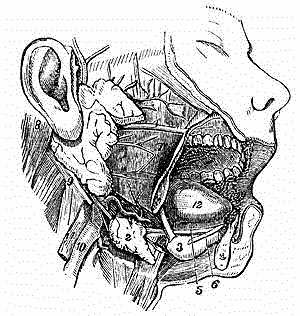
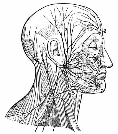 DISTRIBUTION OF THE FACIAL NERVE AND OF THE BRANCHES OF THE CERVICAL PLEXUS.
|
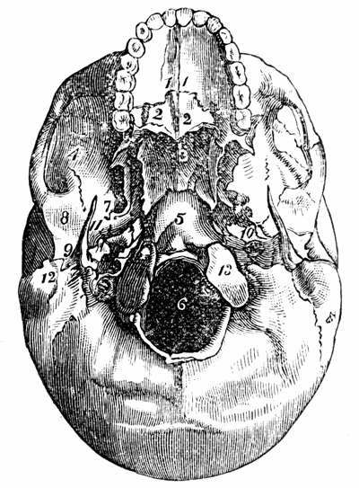 BASE OF THE SKULL.
|
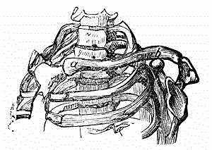
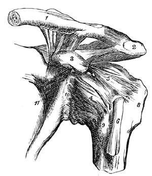 THE LEFT SHOULDER-JOINT AND ITS CONNECTIONS.
|
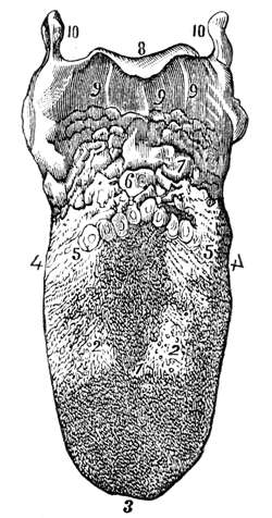 THE UPPER SURFACE OF THE TONGUE, SHOWING THE PAPILLAE.
|